Periapical radiographs are probably the most familiar with images of a few teeth at a time captured on small film cards inserted in the mouth. 810 Periapical periodontitis is a common dental disease ranging from 28 to 12 of teeth.

Periapical Radiography Pocket Dentistry
For this purpose a special technique of periapical radiography was developed by Gordon M.

. Changes to the angulation of the X-ray beam in relation to the teeth and film can help diagnosis and treatment by producing images which provide additional information not always visible on radiographs taken with standard angulations. These patients may include adults with low palatal vaults and children. These X-rays are used to find dental problems below the gum line or in the jaw such as impacted teeth tooth fractures abscesses tumours and bone changes linked to some diseases.
The film is placed parallel to the long axis of the tooth to be radiographed and the central beam of X-ray is directed at right angle to the film and the teeth. A an incorrect vertical angulation. You may be prescribed radiographs that will be used along with your clinical examination.
Periapical radiography is a commonly used intraoral imaging technique in radiology and may be a component of your radiologic examination. A long cone is used to take x-rays with paralleling exposure techniques. Different techniques and instruments are used to drain and decompress large periapical lesions ranging from placing a stainless steel tube into the root canal exhibiting persistent apical exudation 202 204 which is non-surgical decompression to placing polyvinyl or polyethylene tubes through the alveolar mucosa covering the apical lesion which is.
Intraoral periapical radiographs can be produced using two different techniques. Preparing a Patient for the Paralleling Technique. Both techniques have advantages and disadvantages.
Periapical film is held parallel to the long axis of the tooth using film-holding instruments. Machine learning algorithms were also applied in periapical lesion diagnosis using periapical X-ray images panoramic images and cone-beam computer tomography CBCT. By using a filmsensor holder with fixed image receptor and.
Disadvantages to the bisecting technique. Bisecting angle and paralleling. The X-ray head is directed at right angles vertically and horizontally of both the tooth and the image receptor.
Since the slope and curvature of the dental arches and the alveolar processes will not permit the film to be held close to the teeth. The extraoral periapical radiographic technique was performed for both maxillary and mandibular teeth using Newman and Friedman technique2 Maxilla Fig. The central ray is directed to pass at a perpendicular angle to both the tooth and the film.
By using a film sensor holder with still. The image receptor is placed in a holder and positioned in the mouth parallel to the long axis of the tooth under. The bisecting-the-angle technique and the more commonly used long cone paralleling technique.
The X-ray tubehead is then aimed at right angles vertically and horizontally to both the tooth and the image. Assessment of root formation n completion. Demonstration on how to take periapical x-ray using bisecting angle technique About Press Copyright Contact us Creators Advertise Developers Terms Privacy Policy Safety How.
Fitzgerald called as paralleling or long cone technique. Panoramic pan x-rays generate a 5 x 11 or 15 cm x 30 cm wrap-around radiographic image of the patients. A full mouth series of radiographs images the entire dentition and is generally composed of 20 films including 4 bitewing radiographs and 16 periapical radiographs.
1112 Periapical periodontitis is caused by necrotic pulp traumatic or failed endodontic treatment. Periapical X-rays show the entire tooth from the exposed crown to the end of the root and the bones that support the tooth. A full mouth intraoral examination consists of 14 periapical radiographs with two bite-wing films and provides an image of all teeth and related structures.
Parallel technique The image receptor is placed in a holder and placed in the mouth parallel to the longitudinal axis of the tooth under. There are two types of techniques used for periapical radiographs. The film is placed parallel to the long axis.
To take a periapical exposure the hygienist or x-ray technician places a small photosensitive imaging plate coated with phosphorus into a sterile wrapper and inserts it into the patients mouth just like a conventional X-ray film card. The patient was positioned upright with hisher mouth was opened as wide as possible to allow the X-ray beam to pass to the sensor unobstructed from the opposite side of the mouth. The bisecting technique may have to be used for patients unable to accommodate the film positioningdevice used in the paralleling technique.
Single periapical radiographs are often made of individual teeth or groups of teeth to obtain. Periapical radiographs provide important information about the teeth and surrounding bone. Shortened or elongated images.
Periapical x-ray machines are typically mounted on the wall inside each treatment room. For example changes in angulation can be useful to determine the number and curvature of roots and canals to identify superimposed roots and to. Beam of the X-ray not parallel to the proximal surfaces of the teeth.
The paralleling technique results in good quality x-rays with a minimum of distortion and is the most reliable technique for taking periapical x-rays. The paralleling technique is considered to be the best way to take periapical X-rays and when used correctly it should produce reliable images with minimal distortion. Every clinician or dental assistant must be able to take good quality periapical X-rays.
B an incorrect horizontal angulation. C an incorrect incidence point. Periapical radiography is designed to give diagnostic images of the apical portions of teeth and their adjacent tissues.

Periapical Radiography Pocket Dentistry

Periapical Radiography Pocket Dentistry

How Make Periapical X Ray
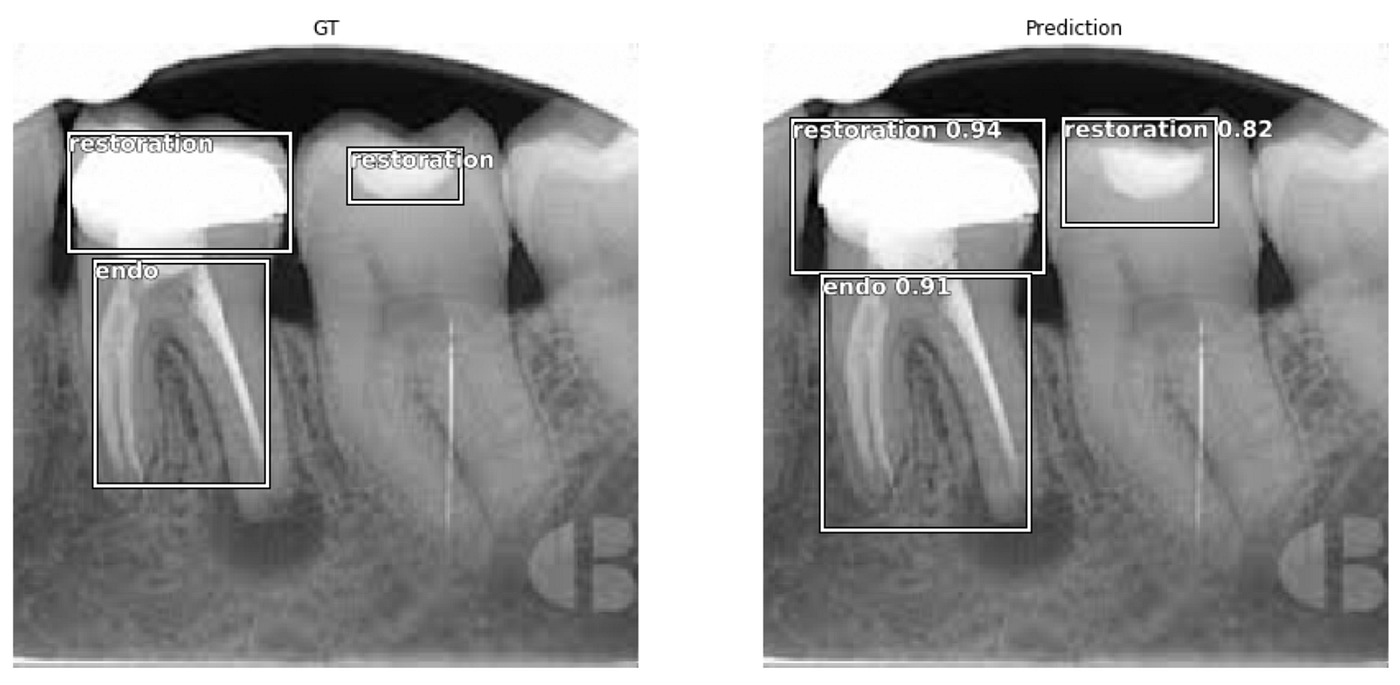
Fastai Object Detection Applied To Dental Periapical X Rays By John Persson Analytics Vidhya Medium
Periapical Radiography Pocket Dentistry

Periapical Radiography Pocket Dentistry

Periapical Radiography Pocket Dentistry

How Make Periapical X Ray
0 comments
Post a Comment